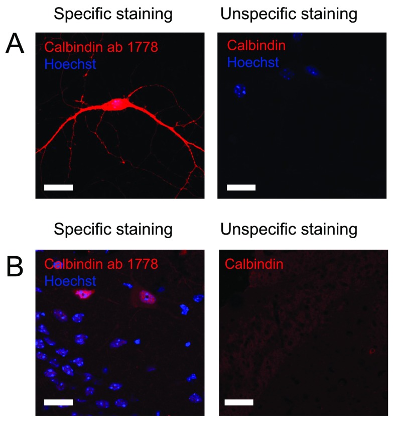
Figure 4. Staining against calbindin.
Figure 4 shows immunostaining against calbindin on A) cultured hippocampal neurons and B) hippocampal tissue. Left pictures shows an example of an immunostaining considered to be specific while right picture shows an example where immunostaining using other primary antibodies did not meet the criteria and therefore was considered unspecific. Scale bar represents 20 µm.

