Figure 5.
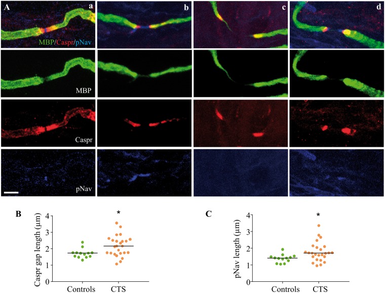
Elongated nodes are characterized by lengthening of the paranodal gap and dispersion of voltage gated sodium channels. (A) Merged images (first row) of nodes of Ranvier triple stained with MBP (green, second row), Caspr (red, third row) and pNav (blue, fourth row) of a control participant (a) and patients with carpal tunnel syndrome (b–d). Panel (a) depicts a normal node of a control participant with clear demarcation of Caspr staining in the paranodes as well as a short band of pNav staining; and (b) shows an elongated node with separation of caspr staining and dispersion of VGSCs within the nodal gap. This was the most frequently identified pattern. We also found occasional elongated nodes with a separation of Caspr staining and absent pNav staining (c). We also identified some heminodes (d), characterized by pNav staining adjacent to the paranodes with a pNav negative gap in the middle of the node. The calibration of 5 µm applies throughout. (B) shows the mean length of the Caspr gap in patients with carpal tunnel syndrome (orange) and control participants (green). This gap was significantly elongated in patients with carpal tunnel syndrome (P = 0.021). (C) The mean length of nodal pNav staining was significantly higher in patients with carpal tunnel syndrome compared to control participants (P = 0.026).

