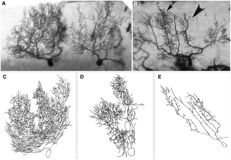
Figure 1.
Reduction in Purkinje cell dendritic complexity in ET. (A and B) Cerebellar cortical sections stained with Golgi Kopsch method, ×2.5. Two adjacent Purkinje cells in a control (A) and one Purkinje cell in an essential tremor case (B). Arrow in B shows an area of relatively preserved dendritic complexity versus arrowhead in B which shows an area of reduced dendritic complexity in the essential tremor case. (C–E) Neurolucida tracings of Purkinje neurons in two controls (C and D) and one representative essential tremor case (E). The neurolucida tracings correspond to different neurons than those shown in A and B.

