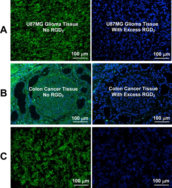
Figure 7.

(A) Selected microscopic images (Magnification: 400×) of living U87MG glioma cells stained with FITC-Galacto-RGD2 in the absence (left) and presence (right) of excess RGD2. (B) Microscopic images (Magnification: 200×) of a tumor slice stained with FITC-Galacto-RGD2 in the absence (left) and presence (right) of excess RGD2. (C) Microscopic images (Magnification: 200×) of the tumor slice (left), which was obtained from a tumor-bearing mouse administered with FITC-Galacto-RGD2 at a dose of 300 μg. Staining with hamster anti-mouse integrin β3 antibody (right) detected with Cy3 conjugated goat anti-hamster antibody (red) was not successful due to blockage of integrin αvβ3 by administration of excess FITC-Galacto-RGD2. (D) Microscopic images (Magnification: 200×) of human colon cancer slice stained with FITC-Galacto-RGD2 in the absence (left) and presence (right) of excess RGD2. Blue color indicates the nuclei stained with DAPI.
