
Figure 8.
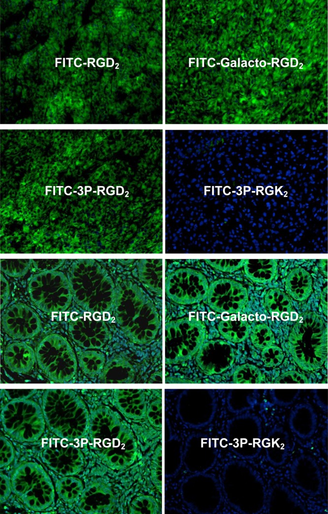
(Top four images) Representative microscopic images of human colon cancer tissues (magnification: 200×) stained with FITC-RGD2, FITC-Galacto-RGD2, FITC-3P-RGD2, and FITC-3P-RGK2. (Bottom four images) Selected microscopic images of U87MG glioma tumor tissues (magnification: 200×) stained with FITC-RGD2, FITC-Galacto-RGD2, FITC-3P-RGD2, and FITC-3P-RGK2. Blue color indicates the presence of nuclei stained with DAPI.