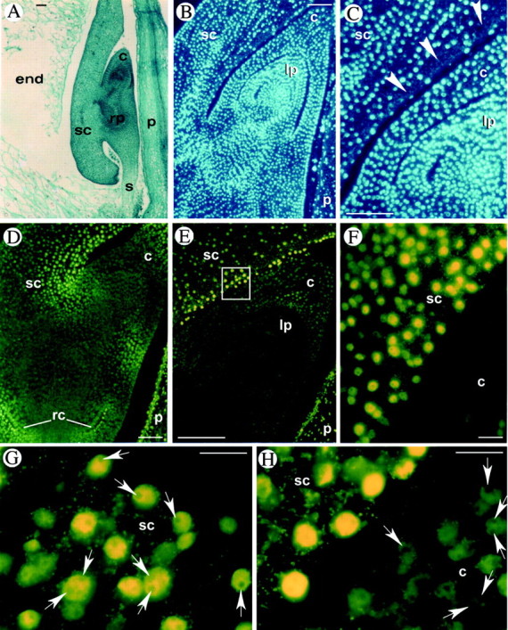
Fig. 1. Programmed cell death in maize embryos at early developmental stages (stages L1–L2). A, Safranin‐fast green staining of a longitudinal section of an embryo at 14 DAP. B and C, DAPI staining of longitudinal sections at 17 DAP showing evidence of nuclear loss (arrowheads) in scutellum layers surrounding the shoot. D–H, In situ detection of DNA fragmentation by the TUNEL procedure (yellow fluorescence on nuclei). TUNEL‐positive nuclei are evident in the scutellum layers surrounding the coleoptile (D–F), in the coleoptile and in the root cap (D). F (enlargement of E) shows the difference between TUNEL‐positive (yellow) and TUNEL‐negative (dark green) nuclei. G, One or two nucleoli (arrows) are present in TUNEL‐positive nuclei of the scutellum at 14 DAP. H, Nucleoli are absent in TUNEL‐positive nuclei and present (arrows) in TUNEL‐negative nuclei at 16 DAP. c, Coleoptile; end, endosperm; lp, leaf primordium; p, pericarp; rc, root cap; rp, root primordium; s, suspensor; sc, scutellum. Bars = 100 µm for A–E and 20 µm for F–H.
