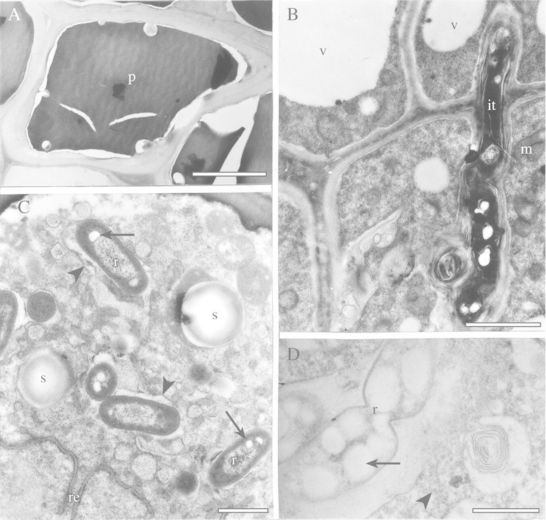
Fig. 3. TEM of endodermal and recently infected cells of angico do cerrado nodules. A, Typical endodermal cell with thick cell wall and polyphenolic compounds (p) in its interior. Bar = 5 µm. B, Infection thread (it) in cells near the nodule meristem. Vacuoles (v) and mitochondria (m) can be seen in the plant cells. Bar = 2 µm. C, The cytoplasm of recently infected cells contains some small starch grains (s) and rough endoplasmic reticulum (re). Rhizobia (r) with small poly‐β‐hydroxybutyrate granules (arrows) are delimited by a membrane (arrowhead). Bar = 1 µm. D, Division of a rhizobium (r), which contains large poly‐β‐hydroxybutyrate granules (arrow) in a symbiosome. Some polyribosomes (arrowhead) can be seen in the host cytoplasm. Bar = 1 µm.
