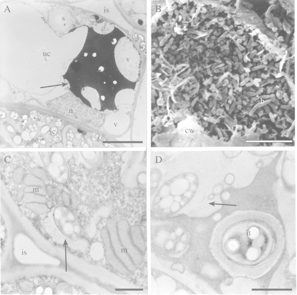
Fig. 4. Ultrastructure of uninfected and infected cells of angico do cerrado nodules. A, TEM of uninfected cell (uc) with vacuoles (v) containing polyphenolic compounds (arrow) occupying part of the cell that has thin peripheral cytoplasm with round starch granules (s). n, Nucleus. An infected cell with symbiosome containing many rhizobia (ic) and intercellular space (is) can also be seen. Bar = 5 µm. B, SEM of mature infected cell with bacteroids (b) occupying almost the entire cell. The remains of the cell wall (cw) are visible. Bar = 10 µm. C, TEM of mature infected cell with dense cytoplasm containing numerous mitochondria (m) near to an intercellular space (is). Note the size of the peribacteroid space (arrow) in the symbiosome. Bar = 1 µm. D, Infection thread (it) in an early senescent infected cell. Note the width of the peribacteroid space (arrow) in symbiosomes. Bar = 1 µm.
