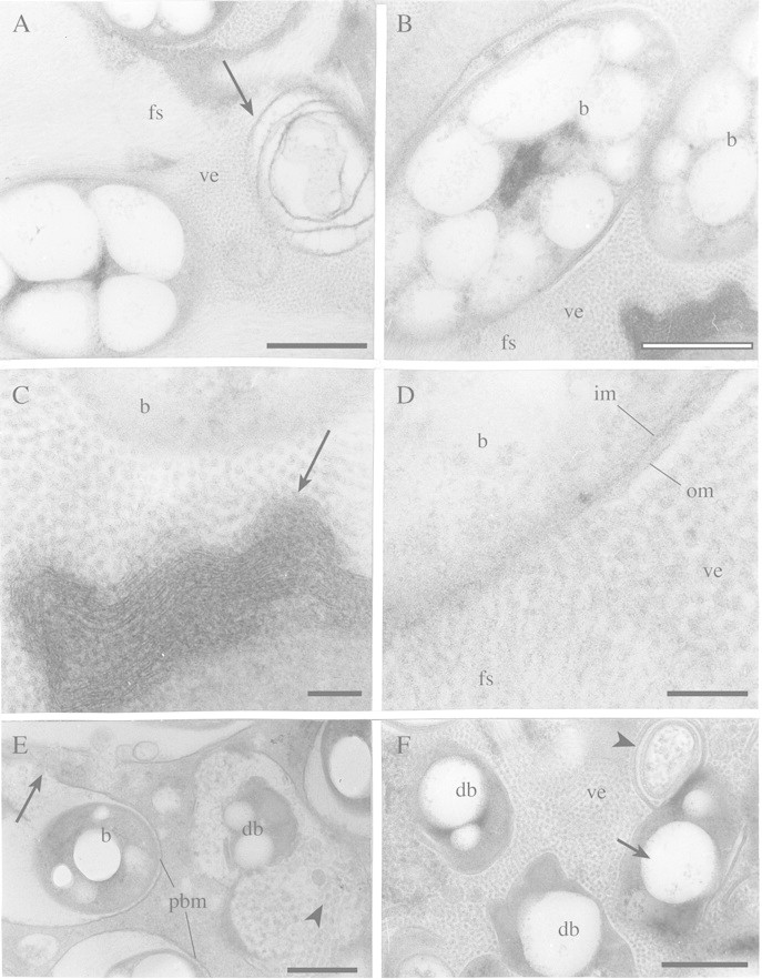
Fig. 5. TEM of infected cells at different stages of senescence of angico do cerrado mature nodule. A, Detail of the peribacteroid space with fibrillar substance (fs) and small vesicles (ve) apparently associated with a reticulum‐like structure (arrow). Bar = 0·5 µm. B, Low magnification of a symbiosome containing bacteroids (b), small vesicles (ve), fibrillar substance (fs) and a different kind of reticulum‐like (arrow) structure (shown in higher magnification in Figs 5C and D). Bar = 0·5 µm. C, Small vesicles may be associated with the reticulum‐like structure (arrow) and with bacteroid (b) cytoplasm lysis in the symbiosome. Bar = 0·1 µm. D, Small vesicles (ve) and fibrillar substance (fs) are in direct contact with bacteroid (b) cytoplasm and may be related to its disorganization. The disappearance of the outer (om) and inner bacteroid membrane (im) can also be seen. Bar = 0·1 µm. E, Bacteroid in advanced stage of degeneration (db) in a symbiosome that contains many vesicles (arrowhead). In other symbiosomes, in earlier stages of degeneration, bacteroids (b) are more intact but the peribacteroid membrane (pbm) that is evident in some cells is disorganized in others (arrow). Bar = 0·5 µm. F, Even later stages of senescence. Degenerating bacteroids (db) still have poly‐β‐hydroxybutyrate granules (arrow). Many vesicles (ve) and membrane‐like structures (arrowhead) can be seen in the host cytoplasm but the peribacteroid membrane is inconspicuous. Bar = 0·5 µm.
