
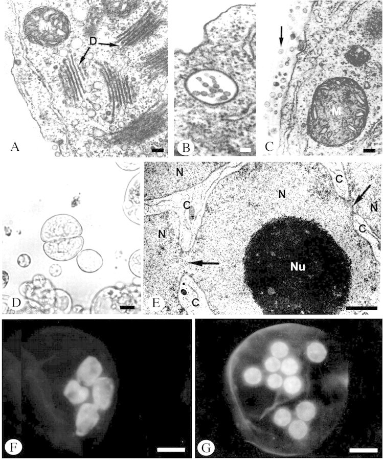
Fig. 5. Changes in the structure and ultrastructure of protoplasts during regeneration of the cell wall, mitotic cell division and amitotic nuclear fragmentation. A–C and E, Material prepared in the standard way for TEM analysis (as in Fig. 4); D, F and G, material in toto: micrographs of live material viewed under a light microscope (D), and in a florescence microscope following DAPI staining (F and G). A, Fragment of protoplast 12 h after isolation showing cluster of dictyosomes (D) and small mitochondrion (M) with clearly visible cristae. B, Multivascular body in protoplast 12 h after isolation. C, Numerous vesicles (marked with arrow) located on the outer side of the protoplast plasmalemma 12 h after isolation. D, Aggregate of two cells formed after division of a mononuclear protoplast 2 d after isolation. E, Constrictions (arrows) in strongly folded nucleus (N) leading to amitotic division. Large nucleolus (Nu) of this nucleus and narrow streaks of cytoplasm (C) are visible between the nuclear fragments. F, Amitotic nuclear divisions in protoplast. G, Nuclei of polynuclear protoplast formed as a result of amitotic divisions. Bars = 0·1 µm (A, B and C), 1 µm (E), 10 µm (D, F and G).