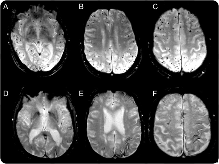
Figure. Representative cases of cerebral microbleed and cortical superficial siderosis predominant cerebral amyloid angiopathy phenotypes.
Axial T2*-weighted gradient echo images in 2 participants, with the first (A–C) showing a high degree of lobar cerebral microbleed burden without cortical superficial siderosis, and the second (D–F) with disseminated cortical superficial siderosis and minimal cerebral microbleed burden.

