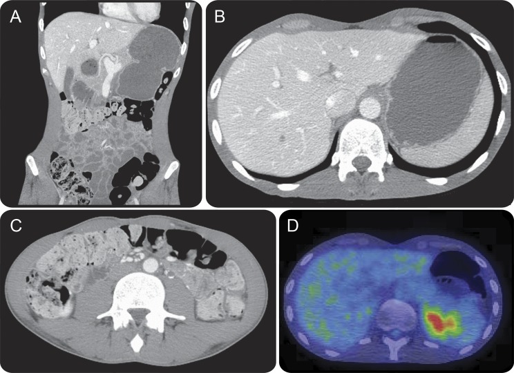
Figure 2. Patient 14: Radiologic evidence of gastrointestinal hypomotility.
The patient defecated at 3-week intervals. Coronal (A), and axial (B and C) CT of the abdomen demonstrates marked gastric distension (A and B), and gross colonic loading with feces (A and C). Fluorodeoxyglucose (FDG)-PET demonstrates retention of food bolus in the gastric fundus secondary to gastroparesis, as well as normal excretion of FDG by the kidneys into the bladder (D).

