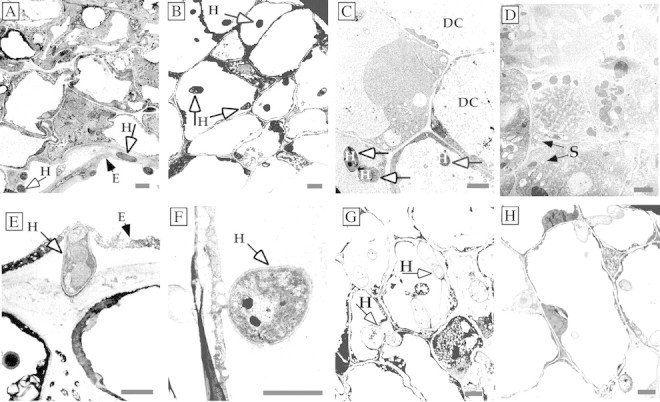
Fig. 5. Tubercles infected with Fusarium oxysporum (A–C) and F. arthrosporioides (D–H) mycelia examined by TEM. Fusarium oxysporum hyphae are inside the epidermal cell wall as well as inside an epidermal cell at 24 h (A and E). Hyphae had penetrated several cell layers of the tubercles by 48 (B and F) and 72 h (G), and the cytoplasm of infected cells and the cytoplasm and cell walls of neighbouring cells were disintegrating by 72 and 96 h, respectively (C and H). Healthy cells with intact organelles in the cytoplasm of the non‐infected control are shown (D). DC, Disintegrated cytoplasm; E, epidermal cell wall; H, hypha; S, starch grains. Bars = 2 µm.
