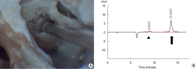
Fig. 1.
Perilymph sampling and quantitative analysis. (A) Demonstration of perilymph sampling at the round window of guinea pig temporal bone on the left side. (B) Glucose (arrowhead) and isosorbide (arrow) peaks detected by high-performance liquid chromatography coupled to refractive index detection.

