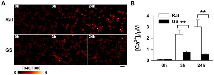
Figure 2. The [Ca2+]i was much lower in ground squirrel neurons 3 and 24 hours after glutamate exposure.

(A) Representative fluorescence ratio images of fura-2. Scale bar: 50 µm. (B) Statistical results show that the relative intracellular calcium concentration ([Ca2+]i) in neurons after glutamate exposure is increased in both ground squirrel and rat neurons. However, the level of the [Ca2+]i increase was much lower in the ground squirrel neurons than in the rat neurons. n = 174–180 neurons, from 3 separate experiments. Two-way ANOVA test with Post hoc Holm-Sidak comparison, P<0.01. Fura-2 AM was used as a calcium indicator.
