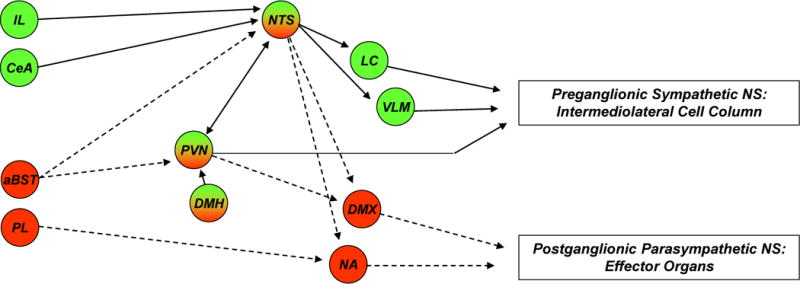
Figure 2. Brain circuitry regulating autonomic stress responses.

Stress-induced pre-autonomic outflow originates in multiple brain areas. Colors denote brain regions that are implicated in sympathetic activation (blue), parasympathetic activation (red) or both (bicolored). The paraventricular nucleus of the hypothalamus (PVN) has substantial projections to both sympathetic and parasympathetic nuclei, including the nucleus of the solitary tract (NTS), dorsal motor nucleus of the vagus nerve (DMX), intermediolateral cell column (IML), locus coeruleus (LC) and ventrolateral medulla (VLM) (latter two not shown for clarity). The rostral VLM, LC, and PVN provide direct innervation of the IML and are thought to initiate sympathetic responses. These NTS in turn receive direct input from neurons in the infralimbic cortex (IL), central amygdala (CeA) and PVN. Other hypothalamic regions, most notably the dorsomedial hypothalamus (DMH), modulate ANS activation via connections with the PVN (and possibly other descending pathways) (see text). Parasympathetic outflow is mediated largely by descending outflow from the DMX and nucleus ambiguous (NA) (colored red) and is under the direct influence of the prelimbic cortex (PL), PVN and possibly other descending relays (see text). Parasympathetic effects of the anterior bed nucleus of the stria terminalis (aBST) are likely mediated by relays in the PVN or the NTS. The anatomical complexity of ANS integration is underscored by the mixing of sympathetic and parasympathetic projection neurons in individual nuclei.
