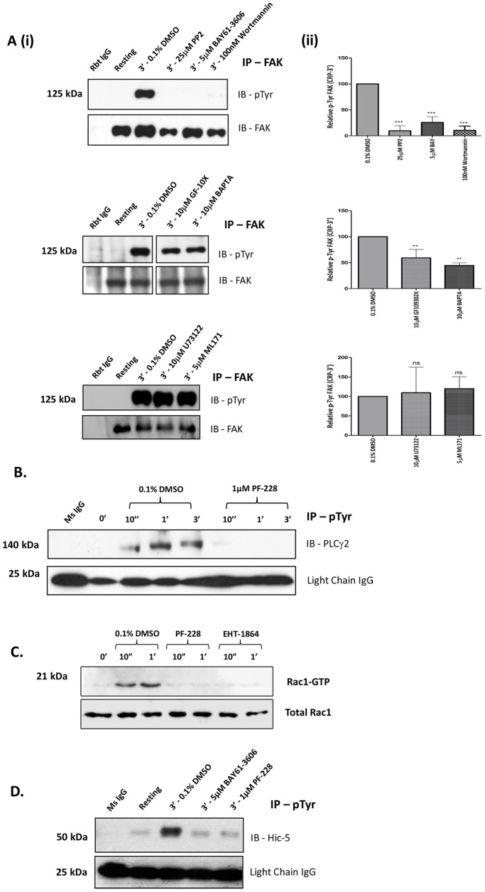
Figure 4. FAK activation within the GPVI pathway.
A(i–ii). Washed human platelets pre-incubated with vehicle control (0.1% DMSO) or inhibitors; 25 µM PP2, 5 µM BAY, 100 nM Wortmannin, 10 µM BAPTA, 10 µM U73122, 10 µM GF109302X and 5 µM ML171 were stimulated with 1 µg/mL CRP for 3 min (with stirring), lysed, immunoprecipitated with anti-FAK and immunoblotted for phosphotyrosine (4G10) and FAK. Representative blots (Ai) and gel densitometry (Aii) presented as Relative pTyr FAK (i.e. pTyr FAK/total FAK) are shown. Data are mean ± SEM (n = 3), (ns) non-significant, **P≤0.01, ***P≤0.001 vs. 0.1% v/v DMSO B. Washed platelets pre-incubated with 0.1% DMSO and PF-228 (1 µM), were stimulated with 1 µg/mL CRP for up to 3 min (with stirring), lysed, immunoprecipitated with anti-phosphotyrosine (4G10) and immunoblotted for PLCγ2. Blots are representative of three independent experiments. C. Washed platelets pre-incubated with 0.1% DMSO, PF-228 (1 µM) and Rac-1 inhibitor, EHT-1864 (50 µM), were stimulated with 1 µg/mL CRP for up to 1 min (with stirring), lysed, subjected to Rac1 GTP ‘pulldown’ analysis and immunoblotted for Rac1 to detect active ‘GTP’ loaded Rac1. Loading controls for total Rac1 levels were subsequently performed using equal sample volumes. D. Washed platelets pre-treated with 0.1% DMSO, BAY (5 µM-included as control) and PF-228 (1 µM) were stimulated with 1 µg/mL CRP for 3 min, lysed, immunoprecipated with anti-phosphotyrosine (4G10) and immunoblotted for Hic-5. IB, immunoblot. Blots are representative of three independent experiments.

