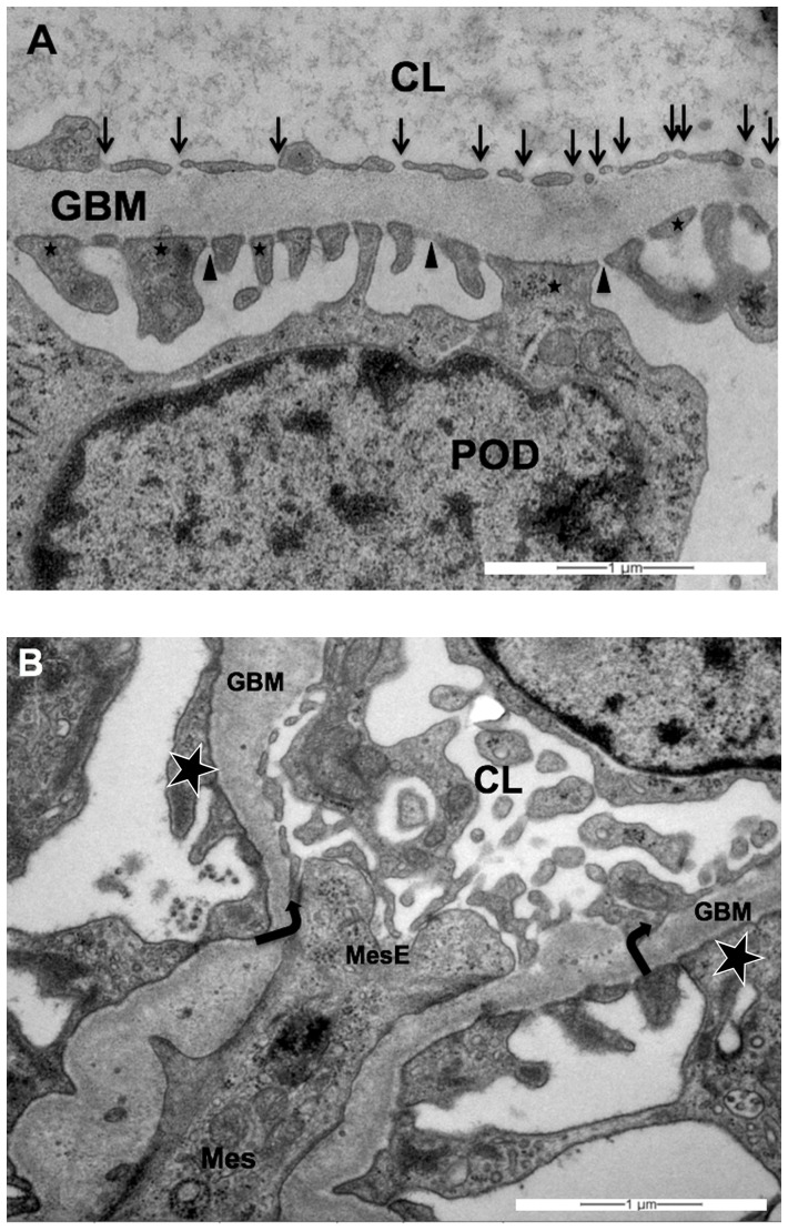
Figure 3. Examples of representative transmission electron micrographs of (A) a fenestrated glomerular endothelium and of (B) peripheral versus mesangial portions of the glomerular endothelium (both one day after aflibercept injection).
(A) Blood lumen on the upper part, urinary space on the lower part of the image. The healthy glomerular filtration barrier consists of three layers [6]: the fenestrated glomerular endothelial cells, the intervening glomerular basement membrane and the podocyte processes and slit diaphragm. GBM = glomerular basement membrane, CL = capillary lumen, POD = podocyte. Arrows mark glomerular endothelial cells fenestrae (note the absence of diaphragm), asterisks mark podocyte foot processes, arrowheads mark podocyte slit diaphragm. (B) At this magnification, podocyte foot processes (asterisks) allow the clear identification of the capillary lumen (CL). In accordance with our definition, the peripheral portion begins where the endothelium and the glomerular endothelial basement membrane (GBM) run approximately parallel (marked by arrows). Arrows mark direction into which peripheral endothelium begins. In between the arrows the mesangium (Mes) and the mesangial portion of the capillary endothelium (MesE) is located. Note that in the mesangial portion there is no GBM adjacent to the fenestrated endothelium so that the described counting method is not applicable and the endothelium does not show the typical single- layered configuration. Magnification ×20000.

