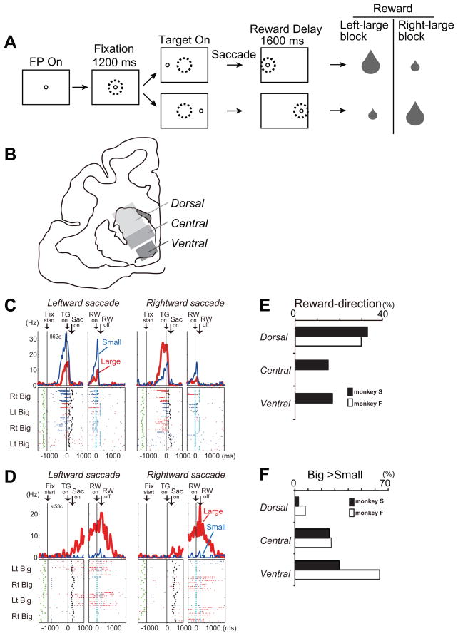
Figure 5.
A. Visually guided saccade task with an asymmetric reward schedule. After the monkey fixated on the FP (fixation point) for 1200 ms, the FP disappeared and a target cue appeared immediately on either the left or right, to which the monkey made a saccade to receive a liquid reward. The dotted circles indicate the direction of gaze. In a block of 20–28 trials (e.g., left-big block), one target position (e.g., left) was associated with a big reward and the other position (e.g., right) was associated with a small reward. The position-reward contingency was then reversed (e.g., right-big block). B. Subdivisions of the primate caudate. C. An example of dorsal caudate neuronal activity showing selective activity associated with the direction of rewarded direction. Note that this neuron showed stronger pre-target activity for the right-big block. The activity in the big- and small-reward trials is shown in red and blue, respectively. The histograms and raster plots are shown in two sections; the left section is aligned to the time of target cue onset (TG on), and the right is aligned to reward onset (RW on). The green dots, the onset of a FP (Fix start); the black dots, saccade onset (Sac on); and the light blue dots, reward onset and offset. D. An example of ventral caudate neuronal activity associated with an expectation of a big reward. E and F. Percentage of neurons that showed reward-direction effect (E) and big-reward preference (F) for each subdivision of the caudate. Modified from Nakamura et al., 2012.

