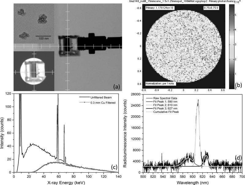
FIG. 1.
(a) Uncollimated, axial projection x-ray image of Y2O3:Eu filled cuvette, abutted to optical bundle fiber for spectra collection during irradiation. Insets: (upper-left) HRTEM image of Y2O3:Eu nanoparticles with 200 nm scale bar and (lower-left) collimated view of same cuvette. (b) Monte Carlo derived phase-space plane at collimator's exit aperture, showing 1.179 × 10¹⁰ x-ray photon flux per mA-sec impinging upon isocenter for mouse phantom studies. (c) Effect upon insertion of 0.3 mm thick Cu filter upon energy spectra, clinically used to prevent patient dosing of low energy, non-therapeutic photons. (d) Typical radioluminescence spectra showing dominant emission peaks at 590 nm, 610 nm, and 627 nm, as well as their corresponding, independent Gaussian curve-fits.

