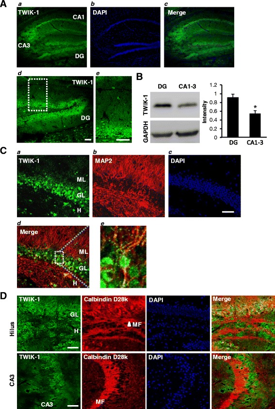
Figure 1.

TWIK-1 is expressed in mouse hippocampal dentate granule cells. (A) Representative fluorescence immunostaining images show that TWIK-1 ( a ) is highly expressed in dentate granular layer and CA1-3 regions. DAPI staining ( b ) indicates the overall hippocampal sub-regions including dentate gyrus and CA1-3. Merged image ( c ) demonstrates co-localization of TWIK-1 with principal cells in all hippocampal sub-regions. (d) Magnified image of dentate gyrus, showing co-localization of TWIK-1 with dentate granule cells. (e) Magnified image of the dotted area indicated in (d). (B) Representative Western Blot data for the expression of TWIK-1 in dentate gyrus and CA1-3 region of the hippocampus (N = 3 mice, P < 0.01, Student’s unpaired t-test). (C) Representative immunostaining images with TWIK-1 (a), MAP2 (b), and DAPI (c). ML, molecular layer; GL, granule layer; H. hilus. Merged TWIK-1 and MAP2 staining image (d) showing that TWIK-1 is co-localized with MAP2 in dendrites of dentate granule cells. High magnification image (e) of dotted rectangle in (d) shows that MAP2-positive proximal dendrites of granule layer cells are co-localized with TWIK-1. Note the presence of TWIK-1 positive cells in the molecular layer (ML) of the dentate gyrus and the hilus (H). (D) Double immunostaining with TWIK-1 (green) and calbindin D28k (red) demonstrates that TWIK-1 is only co-localized with calbindin D28k in the granule layer (GL) but not in the hilus (H) or CA3. DAPI stains neuronal cells in the granule cell layer and CA3 layer. Scale bar, 50 μm. DG: dentate gyrus, GL: granule layer, CA1: cornu ammonis 1, CA3: cornu ammonis 3, ML: dentate molecular layer, H: dentate hilus, MF: mossy fibers.
