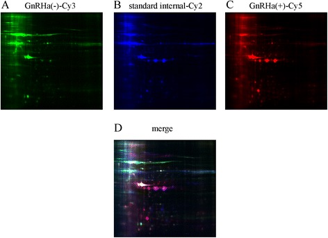
Figure 2.

2D-DIGE of eutopic endometrial proteins. Each individual sample (before and after GnRHa treatment) and internal reference sample was labeled with Cy5, Cy3 and Cy2, respectively, mixed, and separated on a 2D-PAGE gel. The gels were scanned, and Cy5 (A), Cy3 (B) and Cy2 (C) images were obtained from each gel. An overlay of three dye-scanned images was also obtained (D).
