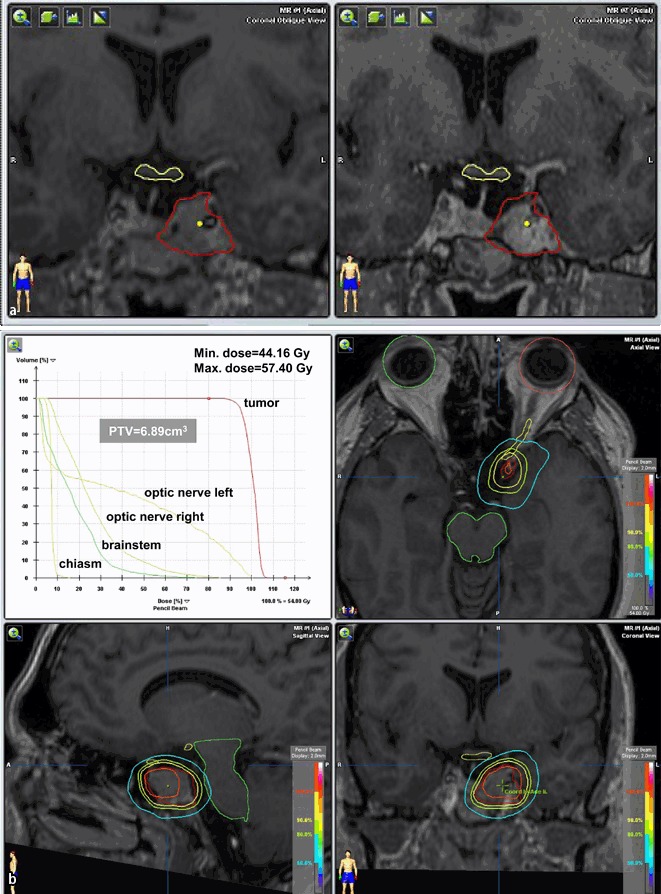
Fig. 2.

a, b MRI of patient (male, 41 years) with tumor progression after 2 transsphenoidal surgeries (the last in 2007), SRT with 5 × 1.8 ad 54 Gy in 2009 (CTV 5.13 ccm). At 3-year follow-up, tumor regression, no visual disorder, hypopituitarism (partial) idem
