
Figure 3.
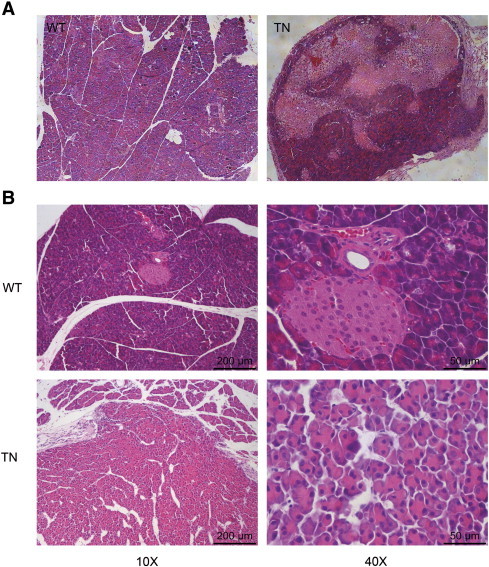
Histology of the pancreas from WT and TN mice. (A) Cross section of normal pancreas (left) and pancreatic neoplasm (right) from representative of 8-month-old wild-type littermates (WT) and Neurog3-Tsc1−/− (TN) mice respectively. (B) Micrograph of normal pancreas (upper) and neoplasm (lower). Hematoxylin and eosin (H&E) staining showed the distinct epithelial neoplasm with cellular and structure resemblance of pancreatic acini.