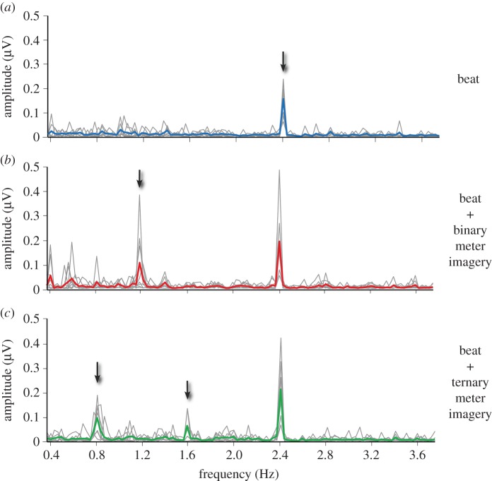
Figure 3.
Beat- and meter-related SS-EPs elicited by the 2.4 Hz auditory beat in (a) the control condition, (b) the binary meter imagery condition and (c) the ternary meter imagery condition. The frequency spectra represent the amplitude of the EEG signal (µV) as a function of frequency, averaged across all scalp electrodes, after applying spectral baseline correction procedure (see [44]). The group-level average frequency spectra are shown using a thick coloured line, while single-subject spectra are shown with grey lines. Note that in all three conditions, the auditory stimulus elicited a clear beat-related SS-EP at f = 2.4 Hz (arrow in a). Also note the emergence of a meter-related SS-EP at 1.2 Hz in the binary meter imagery condition (arrow in b), and at 0.8 Hz and 1.6 Hz in the ternary meter imagery condition (arrows in c). Adapted from [44].

