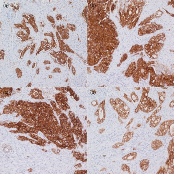
Figure 3.

Staining of human epidermal growth factor receptor 2 (HER2) immunohistochemistry (IHC) 3 + specimen fixed for 8 h (a), 48 h (b), 72 h (c), and 96 h (d). The HER2 IHC 3+ specimen that was fixed for 96 h shows relatively weak staining compared with the other specimens. (Image courtesy of K-M. Kim).
