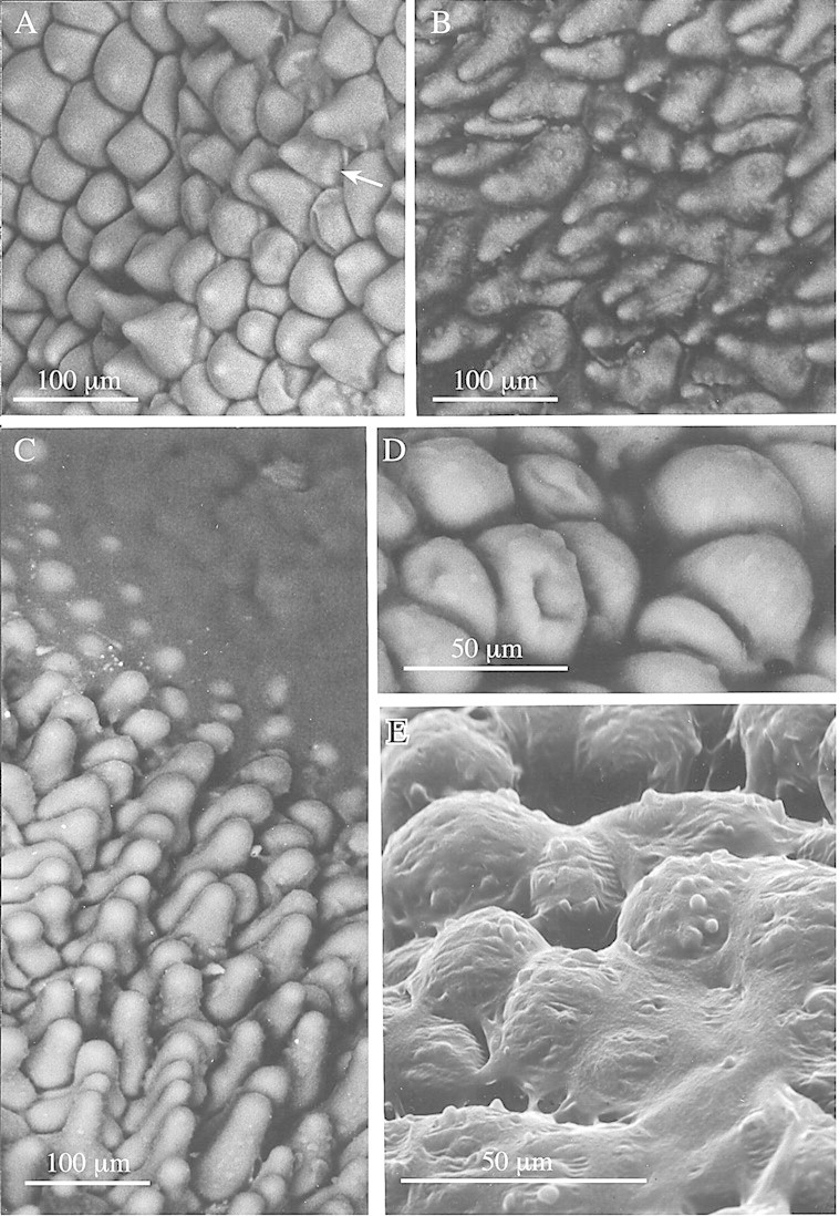
Fig. 1. Low‐vacuum SEMs showing labellar detail of members of the M. acuminata alliance. A, Conical papillae of M. cerifera with some indication of fine, longitudinal sculpturing of the wall (arrow). Bar = 100 µm. B, Conical papillae of M. acuminata showing globules of secretion at tips and along length of papillae. Bar = 100 µm. C, A similar SEM of M. cf. notylioglossa, showing the labellar papillae becoming engulfed by amorphous viscid secretion (top right). Bar = 100 µm. D, Obpyriform papillae from centre of labellum tip of M. cf. notylioglossa. Bar = 50 µm. E, Obpyriform papillae of M. cf. notylioglossa obscured by viscid secretion. Note also the small globules at the tips of the papillae. Bar = 50 µm.
