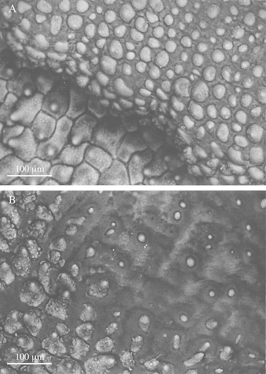
Fig. 2. Low‐vacuum SEMs of labellar surface of M. acuminata. A, Labellar papillae, some showing heavy intercellular deposition of viscid material. Bar = 100 µm. B, A similar SEM, showing papillae tips barely projecting through the thick viscid secretion which, in some cases, has completely engulfed the papillae, resulting in the loss of all topographical detail. Bar = 100 µm.
