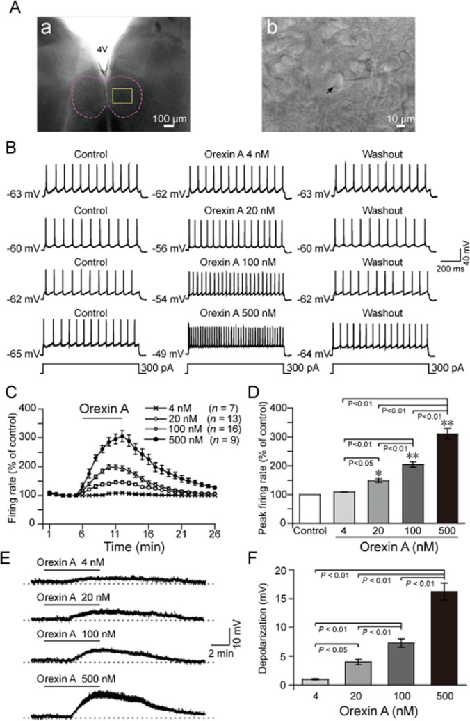
Figure 2.

Orexin A increased the firing rate and decreased the membrane potential of HMNs in a concentration-dependent and reversible manner in neonatal rats. (A) Typical example illustrating the location of the hypoglossal nucleus (a) indicated by a dashed line. (b) Higher magnification of the square area in (a), arrow indicating a hypoglossal motoneuron. The recording electrode is shown entering from the right side of the field. (B) Typical examples illustrating repetitive firing of HMNs before (left), during (middle) and 15 min after (right) perfusion with orexin A at 4 (n = 7), 20 (n = 13), 100 (n = 16) and 500 nM (n = 9), respectively. The evoked depolarizing current steps of these 4 neurons were the same as 300 pA of 1000 ms in duration. (C) Time course of the change in the firing rate induced by orexin A at 4, 20, 100 and 500 nM. (D) Mean peak firing rate (% of control) induced by orexin A. (E) Typical examples illustrating orexin A decreased the membrane potential of HMNs at 4, 20, 100 and 500 nM. (F) Effect of orexin A on the membrane potential of HMNs. Values are means ± SEM. *P < 0.05; **P < 0.01, vs control. 4 V, the fourth ventricle.
