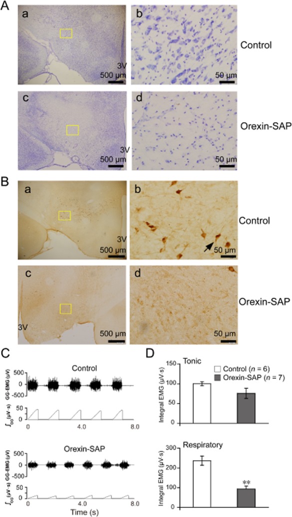
Figure 5.

Lesions of orexin neurons in the bilateral LH after orexin-SAP (400 nl per side, 0.43 mg ml−1) micro-injection decreased the respiratory-related GG-EMG in adult rats. (A) Loss of Nissl bodies in the LH after orexin-SAP treatment. (a) Nissl staining showing cells in the saline-treated rats and (c) the cells in the orexin-SAP lesioned rats. Higher magnification of the square area in a (b) and in c (d). (B) Loss of orexin A-immunoreactive neurons in the LH after orexin-SAP treatment. (a) Anti-orexin A antibody immunohistochemistry staining showing cells indicated by an arrow in the saline-treated rats and (c) the cells in the orexin-SAP lesioned rats. Higher magnification of the square area in a (b) and in c (d). (C) Typical example illustrating orexin neurons lesions decreased the respiratory-related GG-EMG. (D) Changes in the tonic and respiratory-related GG-EMG after orexin-SAP treatment (n = 7) compared with the control (n = 6). Values are means ± SEM. **P < 0.01, vs control. 3 V, the third ventricle; orexin-SAP, orexin-saporin.
