
Figure 5.
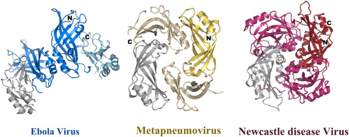
Comparison of dimeric structures of the matrix proteins. Dimeric conformation of the human Metapneumovirus (pdb code 4LP7) matrix protein and of Newcastle disease virus matrix (pdb code 4G1L) in comparison to the Ebola virus VP40 dimer. All three structures have one NTD (grey) in the same orientation. This demonstrates that not only the orientations of the NTD with respect to the CTD are different as shown in Figure 4, but also their mode of dimerization. It should be noted however, that the dimerization mode is quite similar for Metapneumovirus and Newcastle disease virus matrix proteins.