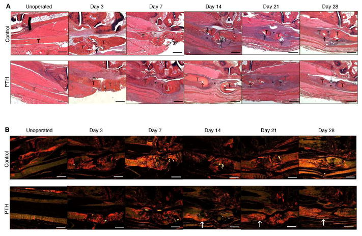
Figure 1.

Representative histologic sections of control and PTH 1-34 un-operated flexor digitorum longus (FDL) tendons and FDL repair tendons at days 3, 7, 14, 21 and 28 post-repair. Tendon ends (T), sutures (↑), and fibroblastic granulation tissue (*) are marked. Sections were stained with (A) alcian blue/hematoxylin and orange G and (B) and picrosirius red illumination by monochromatic polarized light. White arrows indicate areas of increased brightness in PTH 1-34 repairs relative to time-matched controls. Scale bars represent 200 μm. Images are shown at 5X magnification.
