
Fig. 1.
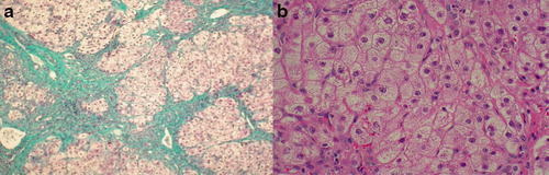
Liver biopsy specimens (2006) age 2 years and 2 months. (a) Wedge liver biopsy (2006): Masson’s trichrome stain shows marked disruption of architecture with extensive fibrosis, portal to portal and central bridging with focal nodules of hepatocyte surrounded by fibrous tissue consistent with early cirrhosis. (b) Wedge liver biopsy: Haematoxylin and eosin stain shows swollen hepatocytes with ballooning degenerative changes in the cytoplasm which have a granular appearance. Stains for glycogen and fat were negative