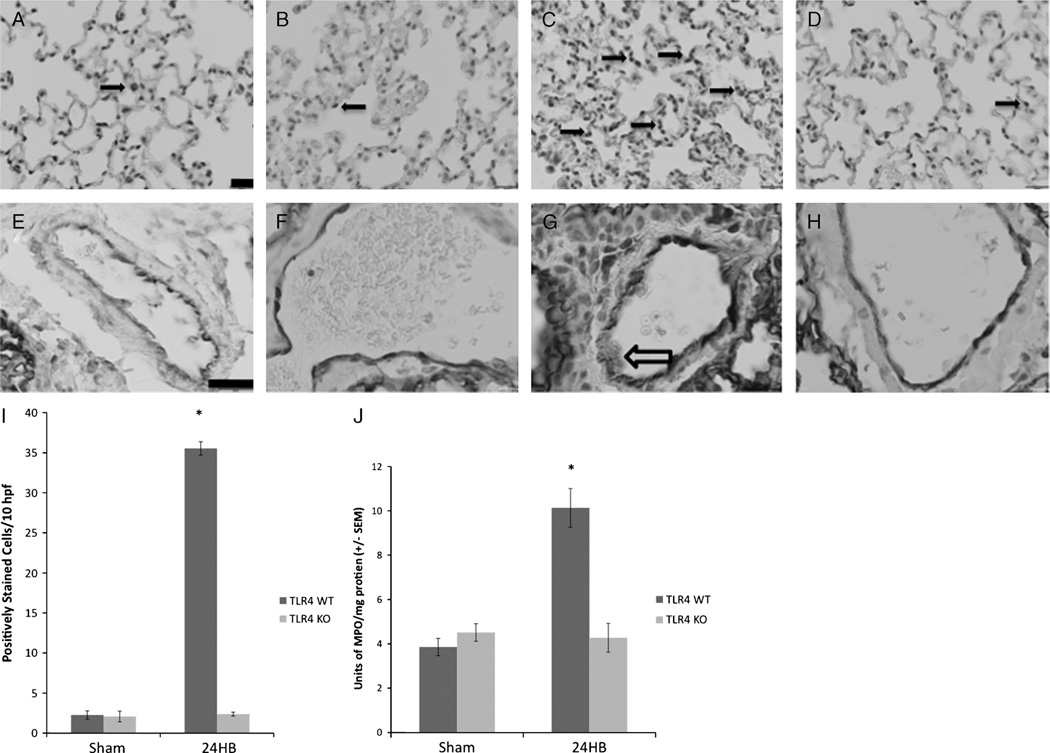
Fig. 6. Lung MPO, ICAM-1 immune staining, and MPO assay.
Immune staining for MPO demonstrates few positively staining cells in sham animal groups (A and B, arrow) but increased in TLR4 WT animals (C) compared with TLR4 KO animals (D). Solid arrows indicate positively staining cells for MPO. Immune staining for ICAM-1 on the endothelial surface of pulmonary arterioles showed minimal staining in both TLR4 WT sham and TLR4 KO sham (E and F, respectively). Deposition of stain on the endothelial surface of pulmonary arterioles in TLR4 WT animals 24 h after burn was more intense than any other group (G, arrow outline). H shows a representative image of ICAM-1 immune staining seen in TLR4 KO animals 24 h after burn, showing similar staining pattern to both sham animal groups. Black bar = 20 µm. A–D, Original magnification ×20. E–H, Original magnification ×60. Myeloperoxidase positively staining cells were counted per 10 high-power fields per experimental group (I). Sections from TLR4 WT animals 24 h after burn had significantly more positively staining cells (*P > 0.05 compared with TLR4 WT sham, TLR4 KO sham, and TLR4 KO 24 h after burn). Enzymatic assay was used to measure MPO levels within pulmonary tissue (J). Myeloperoxidase enzymatic activity was significantly increased in TLR4 WT animals 24 h after burn (*P < 0.005 compared with TLR4 WT sham, TLR4 KO sham, and TLR4 KO animals 24 h after burn). At least four animals were used per group.

