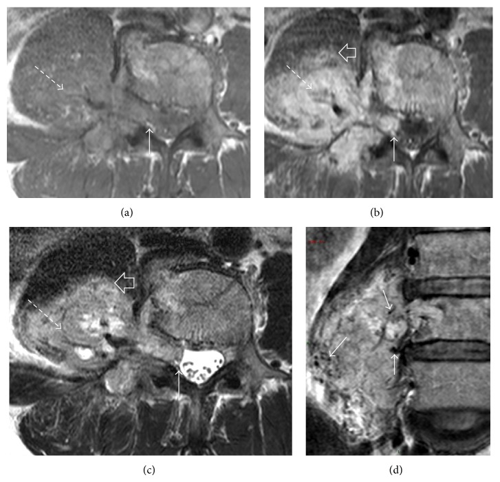
Figure 4.
MRI at L4 lumbar level with axial T1W (a), T1W after contrast (b), and T2W (c) shows a T1-hypointense, T2-heterogenous paraspinal tumour with avid enhancement (dashed arrow). It extends into the moderately widened neural foramen with central displacement of the thecal sac (arrow). The tumour infiltrates the psoas muscle (open arrow). Coronal T2-weighted image (d) demonstrates hypointense tubular structures within the tumour (arrows) compatible with flow voids, indicative of hypervascularity.

