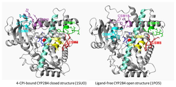
Figure 1.

Open (ligand-free, 1PO5) and closed (4-CPI-bound, 1SUO) structures of CYP2B4 showing CSM 7–11 in the E-H region of the protein. The figures were generated using MOLMOL and Microsoft Publisher files. The CSM 7–11 are labeled and shown in different colors. The I-helix is shown in light green color.
