
Figure 2. Cu-directed anterograde (TGN-to-apical) traffic occurs via TGN-derived vesicles that traverse an EEA1-positive compartment.
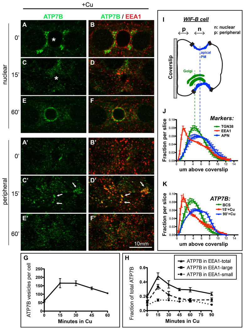
A-B) WIF-B cells were incubated overnight in 10 μM BCS to stage ATP7B at the TGN region then C-D) switched to 10 μM CuCl2 for 15 minutes, or E-F) 10 μM CuCl2 for 60 minutes. Cells were fixed and double-stained with antibodies to ATP7B (green) and EEA1 (red) then imaged by confocal microscopy. A-F) depict single nuclear confocal planes. A'-F' depict single peripheral planes. Renderings of representative stacks from these time points are shown in the top row of Supplementary Figure S4. G) The number of ATP7B vesicles/cell was estimated using 3D objects-based analysis. H) Fraction of ATP7B fluorescence that was coincident with EEA1-positive small and large endosomes. Data shown in G and H represent the mean +/- SEM of at least 4 confocal stacks from a single experiment. I) Schematic cartoon of a WIF-B cell grown on a glass coverslip and rotated 90°. J) Confocal stacks were analyzed by displaying the normalized fractional distribution per slice of various organelle markers, to show the distribution of each marker along the z-plane of the cells. The Cu status of the cells had no effect on the distribution of these markers thus the data shown are pooled with no respect to Cu status, from 7-12 confocal stacks, from at least 2 independent experiments. EEA1-positive endosomes (red) distribute closer to the coverslip (∼2 μm above it) compared to the TGN38 (Golgi, green, ∼5 μm above coverslip) or APN (apical membrane, blue, ∼6 μm above coverslip). K) Cu-induced redistribution of ATP7B into a large basolateral EEA1 endosomes (15 minutes, red profile).