Figure 1.
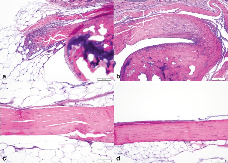
Representative Achilles tendon histologic photographs of db/db (a, c) and wild type (b, d) C57Bl/6J mice. Figure (a) and (b) are at the calcaneus insertion and figures (c) and (d) are of the midsubstance. Note the mild neutrophil infiltration and disruption of the normal architecture in figure a. H&E, bar= 100µ.

