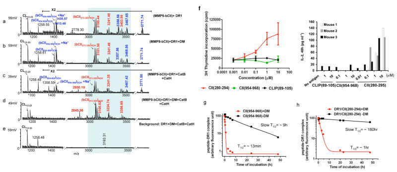
Figure 2. Cathepsins and HLA-DM are necessary for the selection of the immunodominant epitope of type II collagen.
(a–d) Mass spectra of peptides eluted from DR1 containing MMP9-fragmented bCII. e shows the negative control reactions that do not contain the antigen. DR1 used in all experiments shown here (except for in sample d) was pre-incubated with HA(Y308A) to forms short-lived complexes with DR1(DR1/HA(Y308A) complex (T1/2 ~34 min) to induce a peptide-receptive conformation23. The m/z 1258Da peak seen in e is background peptide peak an insect-derived protein that binds to a portion of purified DR1 and is present in most DR1 preparations. The peaks in the shaded area represent post-translationally modified variants of a dominant peptide composed of residues 273–305 of bCII (QTGEPGIAGFKGEQGPKGEPGPAGVQGAPGPAG). Mass species in red represent CII-derived peptides containing the immunodominant core CII(282–289). Non-dominant peptides are shown in blue. Background peptide species are labeled in black. These experiments were repeated more than three times and two cases all three cathepsins were included with similar results. (f) Proliferation of T cells isolated from DR1-transgenic mice immunized with bCII protein in CFA in response to stimulation with, CII(280–294), CII(954–968), or CLIP(89–105) in vitro. Cellular proliferation was measured by [3H] thymidine incorporation (Left panel). The data shown here is a representative result of one mouse out of four individual mice tested. Error bars are defined as SD.
IL-2 ELISA was performed from supernatant collected from in vitro culture of another three individual mice immunized with CII protein/CFA. Cell culture supernatants were removed after 48h culture (Right panel). (g–h) Dissociation assay of fluorescently labeled non-dominant epitope, CII(954–968) and immunodominant epitope CII(280–294) from DR1. Fluorescently labeled CII(954–968)/DR1 complexes (g), or fluorescently labeled CII(280–294)/DR1 complexes (h) were dissociated in the presence of 100μM of unlabeled HA(306–318) at 37°C for the indicated times in the absence (square), or presence of DM (circle). The fluorescence of the labeled complex before dissociation is arbitrarily assigned a value of 100, and fluorescence after dissociation is expressed as a fraction of fluorescence before dissociation. Representative of three dissociation experiments.

