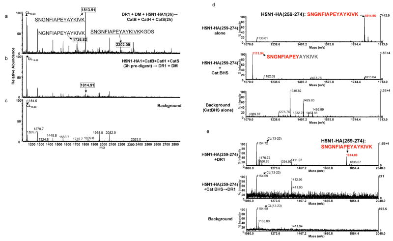
Figure 5. Immunodominant epitope of H5N1-rHA1 is sensitive to the cathepsins.
(a–c) The mass spectra of peptides eluted from DR1 when (a) denatured H5N1-rHA1 (A/Vietnam/1203/2004) was first incubated with DR1 and DM, and then exposed to cathepsins B, H, and S, or (b) when denatured H5N1-rHA1 was first exposed to the cathepsins for 3h and then incubated with DR1 and DM. (c) is the background spectrum. Boxed mass species represent H5N1-rHA1 fragments containing the DR1 restricted immunodominant HA(259–274) epitope. (d–e) mass spectra of the synthetic HA(259–274). (d) Similar to the procedure in Figure 3f, the top panel shows undigested HA(259–274) peptide. Digested peptide with the cathepsins is shown in the middle panel, and the bottom panel shows cathepsins alone as a background control. (e) Mass spectra of peptides eluted from DR1, after 3h incubation of DR1 with HA(259–274) peptide alone (top), HA(259–274) digested with the cathepsins (middle), or the cathepsins alone (bottom). Baculovirus-derived peptide CL(13–23) was detected in every spectra from samples containing DR1. Experiments were repeated three times.

