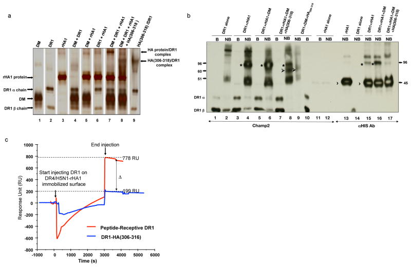
Figure 6. Intact protein antigens form complexes with HLA-DR.
(a–b) SDS-stable complex formation with intact proteins and DR1. Various combinations of DR1, DM, rHA1, and synthetic HA(306–318) peptide were incubated in citrate-phosphate buffer pH 5.0 for 2h at 37°C. Different combinations of proteins and peptides (shown in different lanes) were analyzed by “gentle” SDS-PAGE in which samples are not boiled prior to loading and (a) silver-stained, or (b) western blotted. Polyclonal anti-DR1 serum (CHAMP2) or anti-His antibody was used for detecting protein/DR complexes in (b). Recombinant HA1 protein/DR1 complexes (marked by *) migrated at approximately 96 kD molecular mass. Unbound rHA1 protein migrated at ~45kD (marked by 〉), HA(306–318)/DR1 migrated at around 60kD (marked by ➢), and DR1 is shown at about 50kD. B, boiled, NB, non-boiled.rHA1/DR1 complexes were estimated to migrate at 96 kD molecular mass in both a and b.
(c) Simultaneous binding of two DR alleles to H5N1-rHA1 protein as detected by BIAcore. Biotinylated DR4/HA(Y308A) complex was incubated with denatured H5N1-rHA1 protein for 20min at 37°C in the presence of DM, during which HA(Y308A) would dissociate and be replaced by H5N1-HA1 protein. At the end of the incubation, DR4/H5N1-rHA1 protein complex was injected over the streptavidin coated sensor chip (SA) for 600 seconds and produced 3500 RU, shown as time zero. Then, receptive DR1 (pre-incubated with HA(Y308A) (red), or closed HA(306–318) complex (as control) (blue) were injected over H5N1-rHA1 bound DR4 immobilized surface in the presence of DM for 3000 seconds after which running buffer was injected over the chip to monitor the binding of proteins. Sensograms from both injections are superimposed in the figure for comparison. Negative sensograms observed during the injection are due to changes in buffer conditions between the running buffer and the sample buffer. Binding of the protein of interest to the immobilized protein are shown in the Response Unit (RU) indicating the RU difference between the injection start time versus injection end time.

