Figure 2.

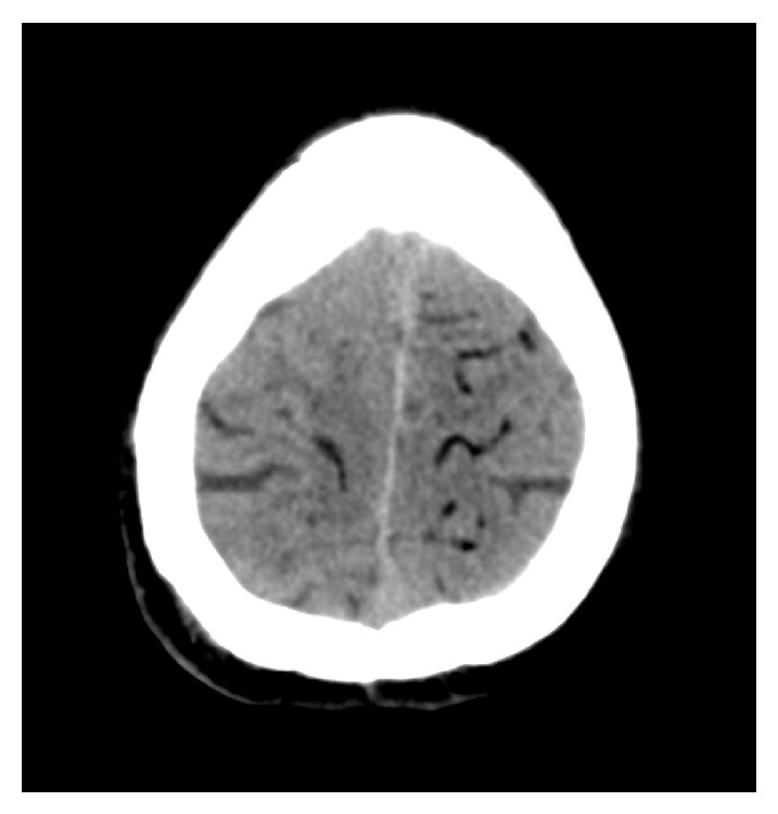
CT image of the brain after lung biopsy with signs of cerebral air embolism, typically visible as subcortical serpentiform formations with negative Hounsfield units.

CT image of the brain after lung biopsy with signs of cerebral air embolism, typically visible as subcortical serpentiform formations with negative Hounsfield units.