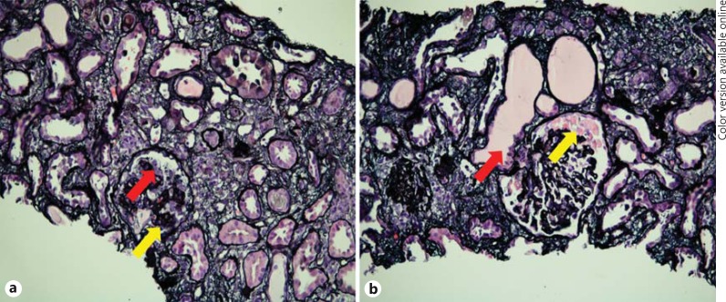
Fig. 1.
Evaluation of the kidney biopsy specimen by light microscopy. a Glomerulus with global collapse of the glomerular tuft (yellow arrow) and podocyte hyperplasia (red arrow). Jones methenamine silver stain. ×200. b Segmentally collapsed glomerulus with prominent protein droplets in visceral epithelial cells (yellow arrow) and acute tubular injury with microcystic dilation of some tubules (red arrow). Jones methenamine silver stain. ×200. Colors refer to the online version only.

