
FIG. 4.
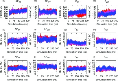
(a)–(l) Number of hydrogen bonds involving residues 15–40 plotted versus time for the Aβ wild type and Iowa mutant. Shown are in the first row the main chain to main chain hydrogen bonding (HB MC MC), in the second row the side chain to side chain hydrogen bonding (HB SC SC), and in the third row the main chain to side chain hydrogen bonding (HB SC MC) for the antiparallel Iowa mutant (APIM), the antiparallel wild-type (APWT), the parallel Iowa mutant (PIM), and the parallel wild type (PWT). The three independent runs are shown in red, blue, and pink.