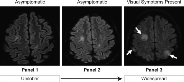
Figure 2.

Representative MRI scans of progression from asymptomatic to symptomatic PML. Asymptomatic PML was diagnosed in a 43-year-old woman with no prior IS use who had previously received interferon beta-1a. Twenty-two months after natalizumab initiation, she had no clinical signs of PML, but MRI showed a hyperintense cortical ribbon on both sides of the superior frontal sulcus (panel 1). Four months later the patient was still asymptomatic, but follow-up imaging showed multilobar lesions and natalizumab was discontinued (panel 2). Six months after first visualization of PML on MRI, PML symptoms, primarily visual, had developed and widespread lesions were present on brain MRI scan (panel 3). Anti-JCV antibody was detected in CSF at this time. MRI, magnetic resonance imaging; PML, progressive multifocal leukoencephalopathy; IS, immunosuppressive; JCV, JC virus; CSF, cerebrospinal fluid.
