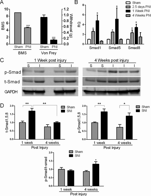
Figure 7.

Muscle Smads are induced and activated in a peripheral nerve injury model. (A) Mice were subjected to sciatic nerve injury (SNI) as described in the Materials and Methods. To determine efficacy of the injury, mice were assessed behaviorally with the Basso mouse scale for locomotion (BMS) 2 days post nerve injury (PNI) and the Von Frey mechanical sensitivity test 1 week PNI (except for the 2.5 day group). There was a significant reduction in locomotion and mechanical threshold to withdraw indicative of nerve injury. ***P < 0.001. (B) Smad mRNA levels were assessed in the gastrocnemius muscle by qRT-PCR at 2.5 days, 1 and 4 weeks post nerve injury (PNI) and compared to a control group which underwent sham surgery. All RNA values were expressed as a fold-change over the 1 week control group. Data are from 3–6 mice in each group. (C) Representative Western blot of muscle extracts from injured (I) or sham controls (S). Antibodies are shown to the left of the blots. (D) Quantitative densitometry of three Western blots representative of 3–6 mice in each test group. For t- and p-Smad quantitation, band densities were adjusted to the loading control (GAPDH) and then expressed as a fold-change over the sham control at 1 week PNI. P-Smad/t-Smad ratios were calculated and expressed as a fold-change over the control group at week 1 PNI. *P < 0.05; **P < 0.005.
