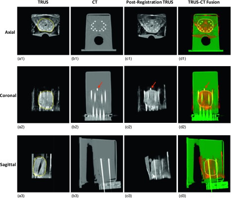
FIG. 4.

3D TRUS–CT registered results of the prostate phantom. (a1)–(a3) are TRUS images in the axial, coronal, and sagittal directions; (b1)–(b3) are CT images in three directions; (c1)–(c3) are the postregistration TRUS images; (d1)–(d3) are the TRUS–CT fusion images, where the prostate volume is transformed from original preregistration TRUS images. The close match between the gold marker (arrows) and catheters in TRUS and CT demonstrates the accuracy of our method.
