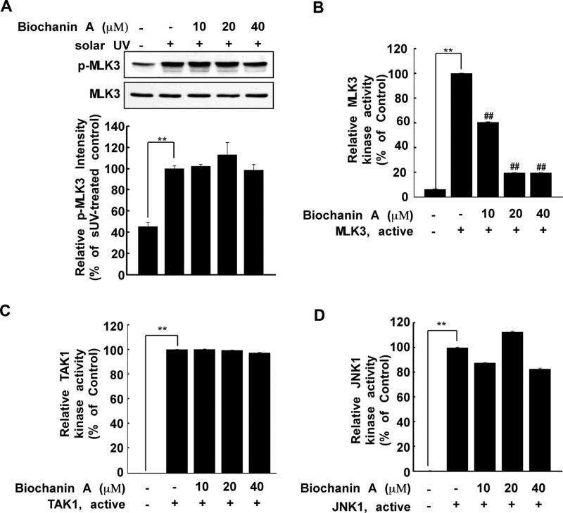
Fig. 3.
Biochanin A inhibits MLK3 kinase activity. (A) After treatment with biochanin A for 1 h, HaCaT cells were treated with sUV. The protein levels of phosphorylated and total MLK3 were measured by Western blotting. Phosphorylated MLK3 and total MLK3 were quantified using the Image J software program. (B) Biochanin A inhibits MLK3 kinase activity. Active MLK3 was incubated with biochanin A at the indicated concentrations for 15 min at 30 °C. MLK3 activity was measured as described in Section 2. (C) and (D) biochanin A does not affect TAK1 (C) or JNK1 (D) kinase activity. Active TAK1 or JNK1 (B) was incubated with biochanin A at the indicated concentrations for 15 min at 30 °C. Kinase activity was measured as described in Section 2. The asterisks (**) indicate a significant difference (p < 0.001) compared to untreated kinase control, and the pound symbols (##) indicate a significant difference (p < 0.001) compared to the untreated kinase control group.

