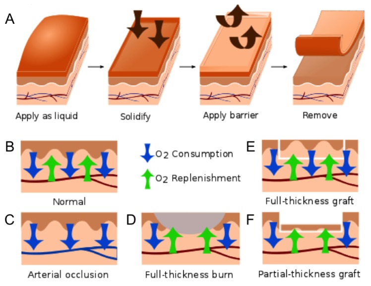
Fig. 1.

A) Schematic diagram showing the application of the pO2-sensing bandage as a liquid, bandage solidification, application of the barrier layer, and bandage removal after pO2 measurement. The supply and consumption of oxygen in tissue balance to yield a measurable tissue oxygenation; B) balanced O2 consumption and replenishment in normal skin; C) decreased surface pO2 during tissue ischemia induced by arterial ligation; D) elevated surface pO2 due to decreased O2 consumption by necrotic tissue in a full-thickness burn; E) unaffected or slightly elevated surface pO2 in a full-thickness skin graft; F) elevated surface pO2 due to decreased O2 consumption at a partial-thickness skin graft, which is composed of non-viable epithelial cells and only a thin layer (30/1000 inch) of dermal cells.
