
Fig. 3.
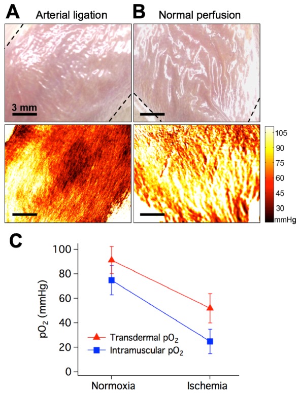
Sensing tissue ischemia in the rat hindlimb. Red indicates lower tissue oxygenation and yellow indicates higher tissue oxygenation. Dashed lines indicate boundaries of bandage-covered region. A) Photograph (top) and tissue oxygenation map (bottom) taken during arterial ligation (lower tissue oxygenation). B) Photograph (top) and tissue oxygenation map (bottom) taken under normal perfusion (higher tissue oxygenation). C) Transdermal pO2 measured by the sensing bandage and intramuscular pO2 measured by the Clark electrode during normal perfusion and ischemia induced by arterial ligation.